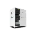
Key Features
- MicroXRF laboratory achieves resolution down to a few micrometers (3-5 µm) with a high-resolution optical system.
- Sensitivity below ppm for quantification down to parts per million (ppm).
- Maximizes throughput and sensitivity with up to 5 different X-ray spectra.
- Allows for overnight scanning without supervision.
- Provides the ability to integrate related techniques such as Raman Spectroscopy.
Detailed Description
Technology & Principles
X-ray fluorescence microscopy (XRF) is a powerful technique with high spatial resolution. This technique uses a finely focused beam that scans the surface of the sample, exciting the atoms within the material. The excitation generates characteristic X-rays, which are then identified to analyze the elemental composition of the sample.
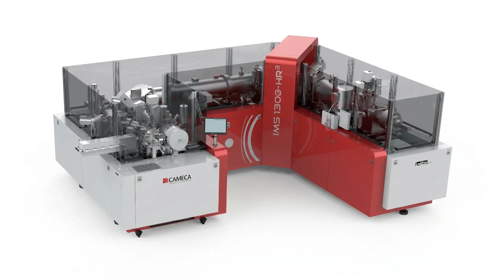
The high-performance capability of the AttoMap-310 is enabled by patented improvements from Sigray, including a patented X-ray source and a high-performance dual parabolic X-ray optical system. The device
provides rapid, non-destructive chemical mapping with resolution down to a few micrometers and a time of up to 5 milliseconds per point.
High-resolution XRF microscopy on the market
X-ray fluorescence microscopy (microXRF) is a technique that provides excellent sensitivity for compositional analysis, with sensitivity typically 1000 times greater than that of electron-based spectroscopy (ppm compared to ppt). The main limitation of laboratory microXRF is the achievable spot size, usually around 20-50 µm. The Sigray AttoMap achieves resolution down to single-digit micrometers (3-5 µm) through the use of Sigray's proprietary X-ray focusing optics. It delivers high performance and produces much smaller spot sizes compared to the multi-channel optical devices used by other laboratory microXRFs.
Sub-ppm sensitivity (Sub-Femtogram)
The AttoMap-200 achieves sensitivity with an absolute detection limit at the sub-Femtogram level and a relative detection limit at the sub-ppm level. This allows the microscope to capture trace element distributions at good throughput.
Energy adjustment capability for high throughput and sensitivity
In contrast to other X-ray sources that are limited to a single X-ray target material, the AttoMap-200 offers software that easily selects up to 5 target materials, including special target materials such as silicon-based and gold-based sources, to ensure optimal sensitivity for a variety of elements.
Large and selective movement range
The robust sample stage of the AttoMap-200 can accommodate large samples or multiple samples (for overnight automated scanning). Samples include: 300mm wafers and large rock core samples.
Integration/ Expansion
The interior space of the AttoMap-200 allows for modifications and upgrades, such as the integration of Raman Spectroscopy.
The AttoMap200 software includes: Single and multi-file analysis, spectrum matching and denoising, implementation of a fundamental parameter (FP) model for quantification without standards, calculation of relative weight percentage using the FP model, spectrum analysis, optical and fluorescence image overlay, ...