

Main features
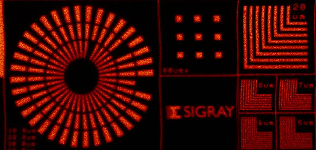
- Laboratory MicroXRF with high resolution in the laboratory achieves a resolution down to a few micrometers (3-5 µm)
- Sensitivity below ppm for quantification down to parts per million (ppm)
- Maximizing performance and sensitivity thanks to a maximum of 5 different X-ray spectra.
- Achieves the ability to image at shallow angles for thin samples (e.g., biological samples) and/or eliminate scattering.
- Allows overnight scanning without the need for supervision. Provides the ability to integrate correlation techniques such as Raman Spectroscopy…
Detailed description
Technology & Principles
X-ray fluorescence microscopy (XRF) is a powerful technique with high spatial resolution. This technique uses a finely focused beam that scans the surface of the sample, exciting the atoms within the material. The excitation generates characteristic X-rays, which are then identified to analyze the elemental composition of the sample.
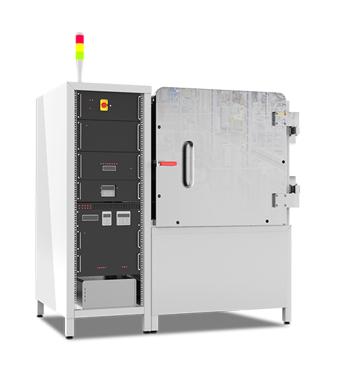
The high-performance capability of AttoMap is enabled by patented improvements from Sigray, including a patented X-ray source and a high-performance dual parabolic X-ray optical system. The device provides rapid, non-destructive chemical mapping with a resolution of a few micrometers and a time down to 5 milliseconds per point.
High-resolution XRF microscope on the market
X-ray fluorescence microscopy (microXRF) is a technique that provides excellent sensitivity for compositional analysis, with sensitivity typically 1000 times greater than that of electron-based spectroscopy (ppm compared to ppt). The main limitation of laboratory microXRF is the achievable spot size, usually around 20-50 µm. The Sigray AttoMap achieves resolution down to single-digit micrometers (3-5 µm) through the use of Sigray's proprietary X-ray focusing optics. It delivers high performance and produces much smaller spot sizes compared to the multi-channel optical devices used by other laboratory microXRFs.
Sub-ppm sensitivity (Sub-Femtogram)
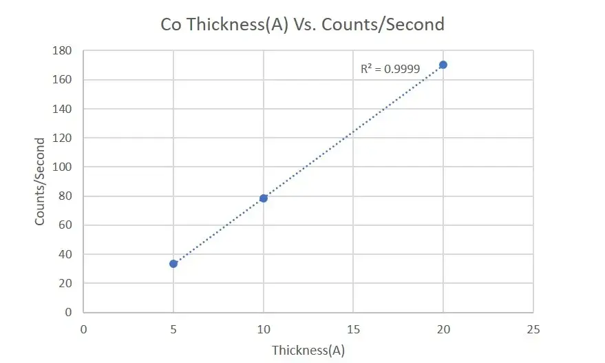
The AttoMap-310 achieves unprecedented sensitivity, with an absolute detection limit at the sub-Femtogram level and a relative detection limit at the sub-ppm level. This allows the microscope to capture trace element distributions at good throughput. The accuracy and speed of the system are why AttoMap has been adopted by leading semiconductor companies to monitor processes related to trace-level impurities.
Energy tunability for high throughput and sensitivity
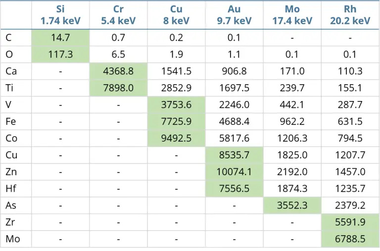
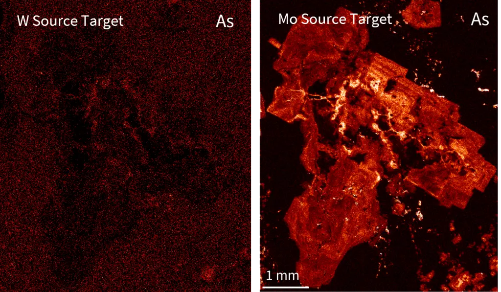
In contrast to other X-ray sources that are limited to a single X-ray target material, the AttoMap-310 offers software that easily selects up to 5 target materials, including special target materials such as silicon-based and gold-based sources, to ensure optimal sensitivity for a variety of elements.
Integration/ Expansion:
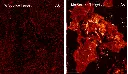
The integrated goniometer allows for the adjustment of various viewing angles, from a perpendicular angle (90 degrees) to an almost oblique angle (3 degrees). For thin samples such as tissue sections or thin films, image quality can be significantly improved at shallower X-ray viewing angles due to the increased X-ray interaction volume and a substantial reduction in background noise. For crystalline samples (e.g., silicon wafers), diffraction peaks can be completely eliminated.