
How does it work?
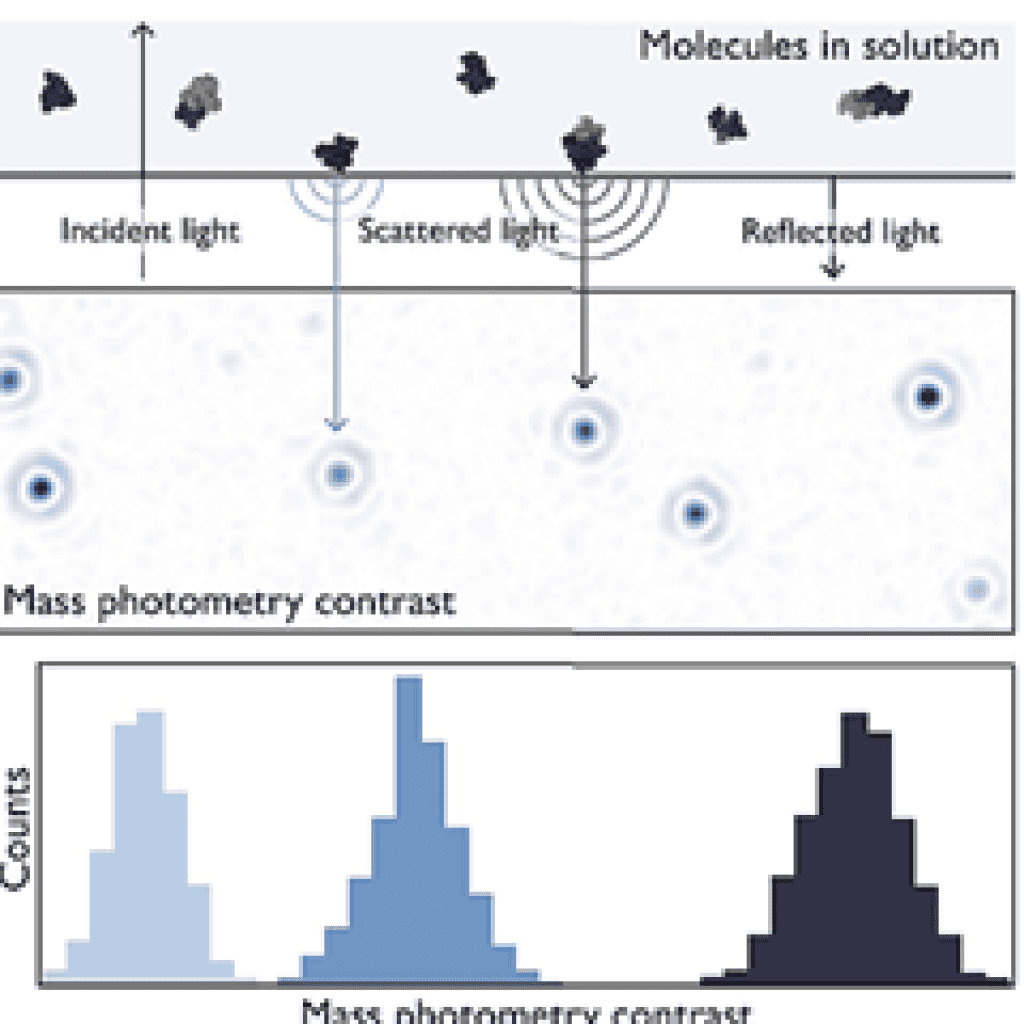
Figure 1: The principle of mass photometry. Light scattered by a molecule attached to the interface measures the interference with the reflected light at that interface. The interference contrast is linearly proportional to the mass.
Light scattered by a particle is linearly proportional to the particle's volume and refractive index. Since the optical properties and protein density change only a few percent, their scattering signal is proportional to their chain mass - allowing for the measurement of individual molecule masses with light, with high accuracy and over a large mass range (Figure 2). The correlation of the scattering signal with mass holds true for many types of biomolecules (glycoproteins, nucleic acids, or lipids), making mass photometry a popular analytical tool for biomolecules in solution.
Benefits of mass photometry
Accurate measurement of natural molecules: In solution, in a range of buffer solutions and compatible with membrane proteins
No labeling, no need for sample modification.
Enter all information under the component in the template.
- Count the molecules.
- Wide block and kinematic bands.
A test provides multiple results
- Homogeneity, structural integrity, and activity
- Fast, simple, minimal sample amount
Applications of the mass photometry method
Purity of the sample and composition
The mass photometry method measures the molecular weight of proteins and protein assemblies in solution with high accuracy. The mass photometry signal scales linearly with the mass of known proteins (as well as other biomolecules) and is independent of their shape. As a result, through simple calibration, mass photometry allows for the measurement of the mass of an unknown with high accuracy (<5 kDa, average 1.9%).

Protein complex - Association and chemical equilibrium
The self-assembly and combination of proteins reinforce their biological function, while protein aggregation can lead to inefficient function, or even toxicity. The mass photometry method provides a unique means to measure the mass distribution of proteins, detecting even very rare quantitative stoichiometries in solution. This provides an excellent insight into the composition of the protein solution and a pathway to test and optimize solution conditions that may affect the oligomerization or synthesis process.
The mass photometry method allows for the determination of the chemical equilibrium distribution of proteins. In a mass photometry experiment, the signal corresponding to individual protein landing events is detected independently of other events.
Single particle detection provides a high dynamic range (3 orders of magnitude in this spectrum) and the ability to detect species with low abundance, such as bovine serum albumin (BSA) tetramers (0.25%). The polydispersity of BSA (monomer 67 kDa) is fundamentally resolved, and the relative amounts of monomer, dimer, trimer, and tetramer can be directly quantified from the number of landing events.

Measurement of complex biomass weight
The mass photometry method is popular for its applicability, as all molecules scatter light, regardless of whether they are based on amino acids, lipids, nucleic acids, carbohydrates, or other building blocks. This means that not only molecules with simple compositions but also molecules that include multiple layers of biomolecules (e.g., glycoproteins, detergent-solubilized membrane proteins, protein-DNA complexes) can be measured with precise mass measurements using the mass photometry method.
The differences in molecular mass of heterogeneous biomolecules, such as lipid nanodiscs, can be distinguished and have high reproducibility. Measurements using alternative techniques - size exclusion chromatography, nuclear magnetic resonance spectroscopy, dynamic light scattering, and natural mass spectrometry - return mass values that differ by about 30% (the orange lines). The mass photometry method can assign accurate mass values to nanodiscs with varying protein belt components and lipid composition. Analyzing lipid bilayer simulations, such as nanodiscs, is a key capability for studies on the structure and function of membrane proteins.

Protein-protein interaction
The function of proteins, and their malfunction in disease, is governed by their interactions with themselves and with other biomolecules. The mass photometry method provides a unique and powerful new means to quantify these interactions in terms of physically meaningful quantities, such as dissociation constants and free energy. This opens the door to measuring the manipulation of these interactions in drug development.
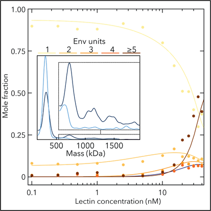
Complex protein-protein interactions can be quantified using mass photometry. The Env glycoproteins of the HIV virus are targeted by antiviral viruses that bind to Env units or cross-link. At low concentrations of the lecturer, Env is primarily a monomer. At higher concentrations, higher-order Env units are detected and found to include bound lecturers. From the titration curve and the relative amount of the Env-speechin complex, the binding affinity of each interaction can be quantified providing key information to understand antiviral mechanisms.
Refeyn One MP - A new era in the analysis of biomolecules
A revolutionary approach to biological analysis - Refeyn One MP marks the beginning of a new era in the analysis of biomolecules. Using mass photometry technology, OneMP allows for the measurement of the mass of individual molecules directly in solution. This unique technology detects a portion of light scattered by individual molecules at the observation interface and uses this signal not only to count molecules in solution but also to measure their mass. All of this is done by measuring each molecule in a series of individual buffer solutions, without the need for labeling.